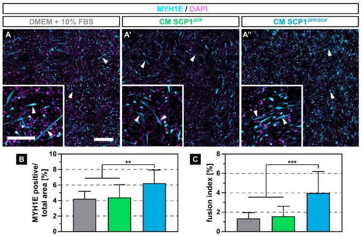
Figure 3.
Immunohistochemistry against myosin heavy chain 1E (MYH1E) (turquoise) after 5 days of myogenic differentiation showed newly fused myotubes in all culture conditions (A–A’’, arrowheads). However, myoblasts that were exposed to conditioned medium (CM) derived from SCP1GFP/SCX cells showed an increased fusion, when compared to C2C12 in NM or CM of SCP1GFP cells (A-A’’). Quantification of the MYH1E positive area (B) and the fusion index (C) validated the microscopic appearance. Scale bars: 500 µm (overview), 200 µm (insert). Bar plots represent the mean and SD. ** equals p ≤ 0.01, *** equals p ≤ 0.001. Five randomly selected pictures of three independent experiments were analyzed.

