Figure 4.
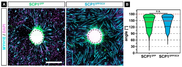
Immunohistochemistry against MYH1E (turquoise) after 7 days of myogenic differentiation in co-culture with SCP1GFP or SCP1GFP/SCX cell pellets revealed that most of the newly formed myofibers were orientated towards the middle of the cell pellets (A). However, the quantification of the orientation did not show any significant difference between both groups (B). Vertical lines in violin plots represent the median (red) and quartiles (black). Red dashed lines indicate a perfect orientation towards the center of the pellet. n.s.: not significant. Scale bar: 500 µm.

