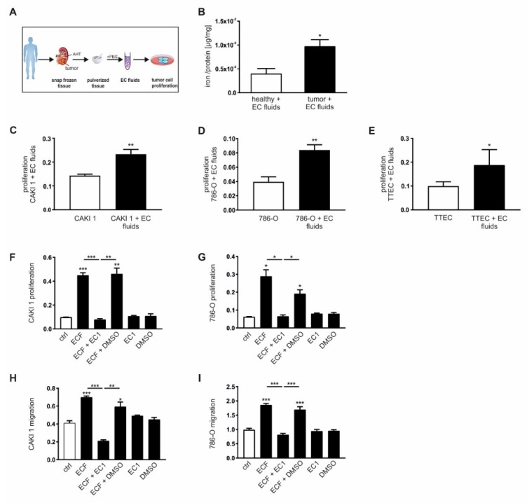
Figure 4.
Extracellular iron induces proliferation and migration of tumor cells in vitro. (A) Schematic overview of how to generate extracellular (EC) fluids (ECF) from primary human renal tumor and adjacent healthy tissue. (B) Iron load measured by AAS relative to the total protein amount of EC fluids of ccRCC tissue compared to adjacent healthy renal tissue (n = 8). Proliferation of (C) CAKI-1 (n = 7), (D) 786-O (n = 8), and (E) primary human tumor tubular epithelial cells (TTEC) upon stimulation with EC fluids in vitro measured with the xCELLigence system (n = 8). Proliferation of (F) CAKI 1 (n = 4) and (G) 786-O (n = 4) cells as well as migration of (H) CAKI 1 (n = 4) and (I) 786-O cells (n = 4) upon stimulation with EC fluids in the presence or absence of an extracellular chelator (EC1, 100µM) or dimethyl sulfoxide (DMSO) as negative control measured with the xCELLigence system. Graphs are displayed as means ± SEM with * p < 0.05, ** p < 0.01, *** p < 0.001.

