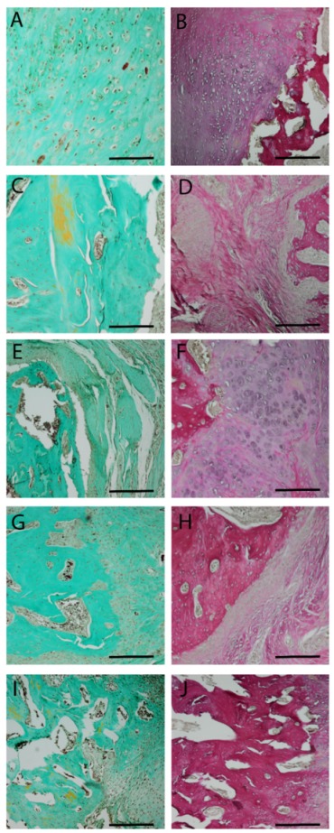
Figure 6.

Masson–Goldner (first column) and Elastica van Gieson (second column) staining 8 weeks after surgery. Central parts of the fracture are shown. Group 1—no implant—(A,B); 2—PLA cylinder alone—(C,D); 3—PLA cylinder + collagen type I—(E,F); 4—PLA cylinder + collagen type I + SDF-1—(G,H); 5—PLA cylinder + collagen type I + BMP-7—(I,J). Scale bars: 1000 µm.
