Figure 5.
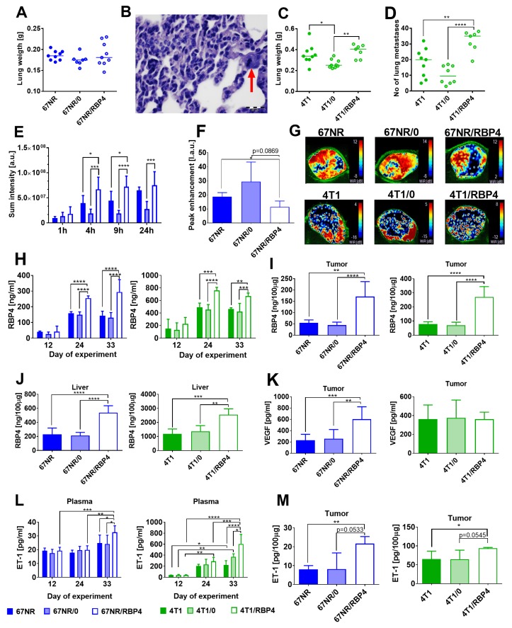
The effect of RBP4 overexpression on metastatic potential and angiogenesis of 67NR and 4T1 tumors. (A) Lung weight of 67NR tumor-bearing mice (N = 7–9) and (B) microphotograph of lung metastasis in mice bearing 67NR/RBP4 cells. Red arrow indicates epithelial cell with mitotic spindle. (C) Lung weight and (D) number of lung metastatic foci in mice bearing 4T1/RBP4 tumors (N = 7–9). (E) Blood vessel permeability in 67NR/RBP4 tumors (N = 4). (F) Peak enhancement in tumor tissue of mice bearing 67NR/RBP4 tumors (N = 3–4). (G) Representative pictures of wash in rate parameter. Concentration of RBP4 protein in (H) plasma (N = 3–5), (I) tumor tissue (N = 5), and (J) liver (N = 5). (K) VEGF in tumor tissue (N = 6–9). Concentration of endothelin-1 (ET-1) in (L) plasma (N = 3–4) and (M) tumor tissue (N = 3–4). Data presented as mean ± SD or data for individual measurements (Figures (A), (C), and (D)). Statistical analysis: Tukey’s multiple comparison test. * p < 0.05, ** p < 0.01, *** p < 0.001, **** p < 0.0001.

