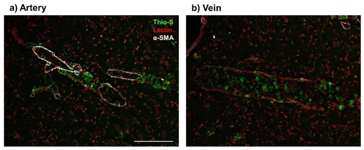
Figure 1.
Presence of venular amyloid in 16-month old TgF344-AD rats. Immunofluorescence imaging of vascular amyloid deposition, stained by Thioflavin S (Thio-S, green), in cortical penetrating vessels (Lectin, red). Amyloid beta peptide (Aβ) deposits present a ‘double-barreling’ morphology, forming cyclical rings surrounding the arteries (a). Venular Aβ is present as smaller, globular deposits along the veins (b). Arteries were distinguished from veins by presence of alpha smooth muscle actin (α-SMA, white). Scale bar = 100 μm.

