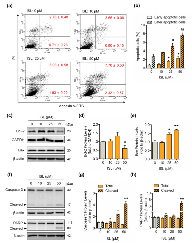
Figure 3.
ISL induced cell apoptosis by upregulating apoptotic protein expression in MDA-MB-231 cells. (a) MDA-MB-231 cells were treated with ISL for 48 h. Cells were stained with propidium iodide and annexin V fluorescein isothiocyanate (FITC), and the apoptosis rates were analyzed by flow cytometry. (b) The quantitative data of apoptotic cell death in early and late phases are shown. (c) After treatment as indicated above, the anti- and proapoptotic proteins were monitored using Western blotting in MDA-MB-231 cells. (d,e) Each target protein was normalized to GAPDH or β-actin expression. (f) The expression of caspase-3 and its downstream molecule, PAPR, was monitored using Western blotting in MDA-MB-231 cells. (g,h) Each target protein was normalized to β-actin expression. Data are represented as mean ± SEM (n = 3). * p < 0.05, ** p < 0.01 compared with the control group. + p < 0.05 compared with the early apoptotic phase of the control group. # p < 0.05, ## p < 0.01 compared with the later apoptotic phase of the control group.

