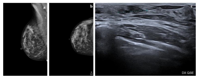
Figure 1.
Radiologic features of a low-grade ductal carcinoma in situ (DCIS) lesion. (a,b) Mammograms show a heterogeneously dense right breast. In the upper outer quadrant (UOQ) there is a small oval opacity with obscured margins; (c) ultrasound shows an irregular mass with indistinct margins, hypoechoic echo pattern without posterior features.

