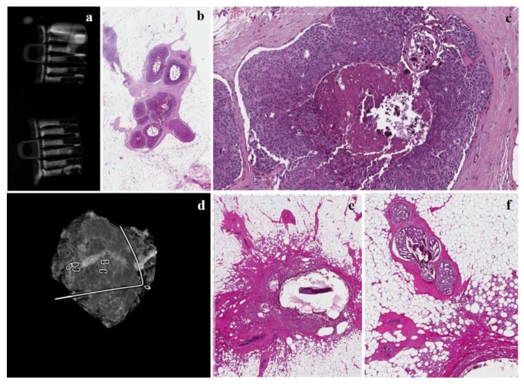
Figure 3.
The same case as Figure 3. (a) Radiograph during localization of the microcalcification in the samples collected in touch-free collection chambers using the Mammotome Revolve 10 gauge biopsy system that reveals numerous microcalcifications in the cores; (b,c) mammotome biopsy shows a high-grade DCIS with central comedonecrosis at low- and high-magnification; (d) mammogram revealing successful retrieval of a cluster of pleomorphic calcifications with prior localization and subsequent surgery; (e) inflammatory reaction around the previously placed clips; (f) a residual focus of cribriform carcinoma in situwith central microcalcifications.

