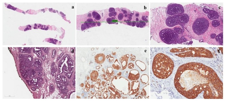
Figure 4.
Features of a low-grade DCIS. (a) Low-grade DCIS in a core needle biopsy; (b,c) intermediate and high-magnification of the lesion showing a cribriform growth pattern; (d) DCIS with negative margins, but the distance between neoplasia and inked margin is <2 mm; (e) ER positivity in a low-grade DCIS; Her-2 positivity (score 3+) in a low-grade DCIS.

