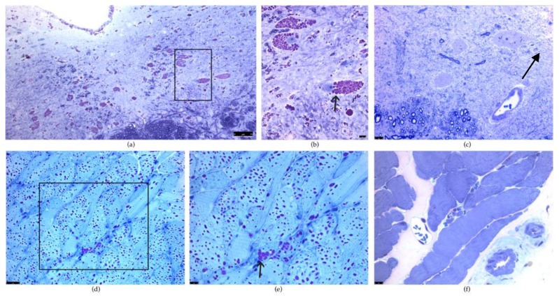
Figure 2.
Periodic acid–Schiff (PAS) staining of the hypoglossal motor neurons and the tongue in Gaa−/− and wild type (WT) mice. (a,b): PAS staining is positive in the hypoglossal motor nucleus in Gaa−/− mice. (b) is a higher magnification of the boxed region (a) and illustrates PAS positive vacuoles indicative of glycogen-filled lysosomes in the hypoglossal motor neurons. Note the disruption in the architecture of the motor neuron resulting in displacement of the nuclei (arrow). (c) illustrates PAS staining in a WT mouse at the same magnification as (b). Note the lack of positive PAS staining in the XII motor neuron and the centrally placed nuclei (arrow). (d,e): PAS staining of a Gaa−/− tongue illustrates vacuoles filled with PAS positive glycogen (arrow). (d) is a higher magnification of the boxed region (d). (f): PAS staining of a WT tongue shows striated muscle with no evidence of PAS positive vacuoles. (f) is the same magnification as (e). Scale bars lower right in (a,b); lower left corner in (c,d,e,f). The scale bar denotes 7µm in (a), 10µm in (b,c,e,f), and 25µm in (d).

