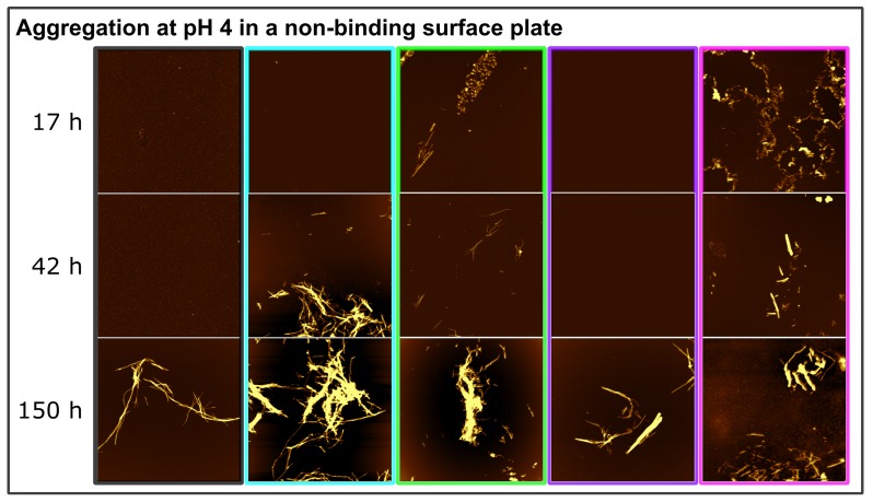
Figure 6.
Time-resolved AFM height images of -synuclein aggregation at pH 4 in a non-binding surface plate without glass beads. The colors of the frame correspond to the conditions (Figure 3): control (black frame), EGCG (1:1) (cyan frame), EGCG (1:5) (green frame), EGCGox (1:1) (purple frame), and EGCGox (1:5) (magenta frame). The image scale is 5 × 5 μM. The color range of the image represents the height range from −5 to 20 nm.

