Figure 7.
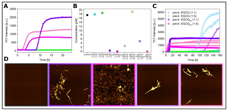
Aggregation kinetics of -synuclein at pH 4 in a non-binding surface plate under quiescent conditions in the absence of glass beads. The fibril formation was monitored in the presence and absence of EGCG or EGCGox and in wells that were pre-treated with EGCG-solutions (A) and the corresponding concentration measurement by Fluidity One after 160 h (B) with AFM height images (D) of the aggregation products of -synuclein (black frame) in the presence of EGCGox (1:1) (purple frame) and (1:5) (magenta frame), in the pre-treated wells with EGCGox (1:1) (light purple frame) and (1:5) (light magenta frame), and the overview of the three replicates per condition (C). The image scale is 5 × 5 μM. The color range represents the height from −3 to 12 nm.

