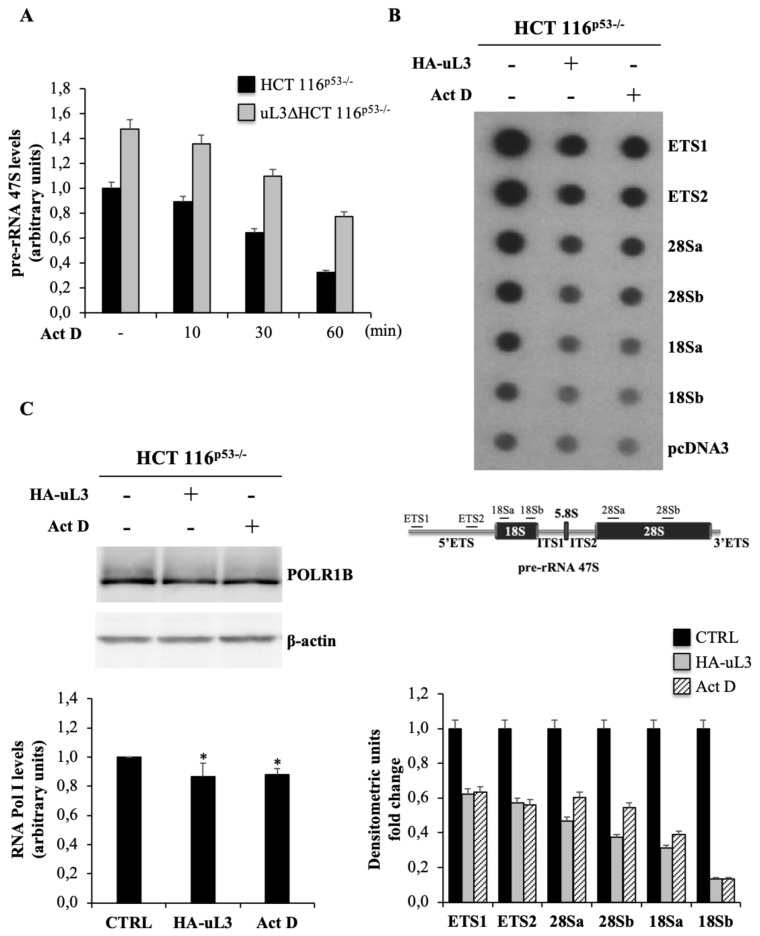
Figure 2.
uL3 status affects 47S pre-rRNA synthesis and stability. (A) Total RNA from HCT 116p53−/− and uL3ΔHCT 116p53−/− cells, treated with Act D 5 nM for 0, 10, 30 and 60 min was subjected to RT-qPCR with primers specific for 5′ETS region of 47S pre-rRNA and β-actin mRNA (Table 1). Quantification of signals is shown. Bars represent the mean of triplicate experiments; error bars represent the standard deviation. (B) Nuclear run-on assay. 20 μg of plasmid DNA including sequences of two regions of 5′ETS, two regions of 28S, two regions of 18S and plasmid pcDNA3 were spotted on membrane and incubated with 32P-labeled RNA from untreated HCT 116p53−/−, HCT 116p53−/− cells transfected with 1 µg pHA-uL3 and HCT 116p53−/− cells treated with Act D. The average of two signals normalized for pHA-uL3 is reported in the bar graph in the lower panel. A schematic diagram of 47S pre-rRNA indicating the regions used in nuclear run-on assay is shown in the lower panel. Full-length blot is shown in Figure S5. (C) Total protein extracts from HCT 116p53−/− cells untransfected or transfected with 1µg pHA-uL3 and HCT 116p53−/− cells treated with Act D were analyzed by Western blot with the indicated antibodies. Full-length blots are shown in Figure S6. Quantification of signals is shown. Bars represent the mean of triplicate experiments; error bars represent the standard deviation. * p < 0.05 vs. HCT 116p53−/− cells set at 1.

