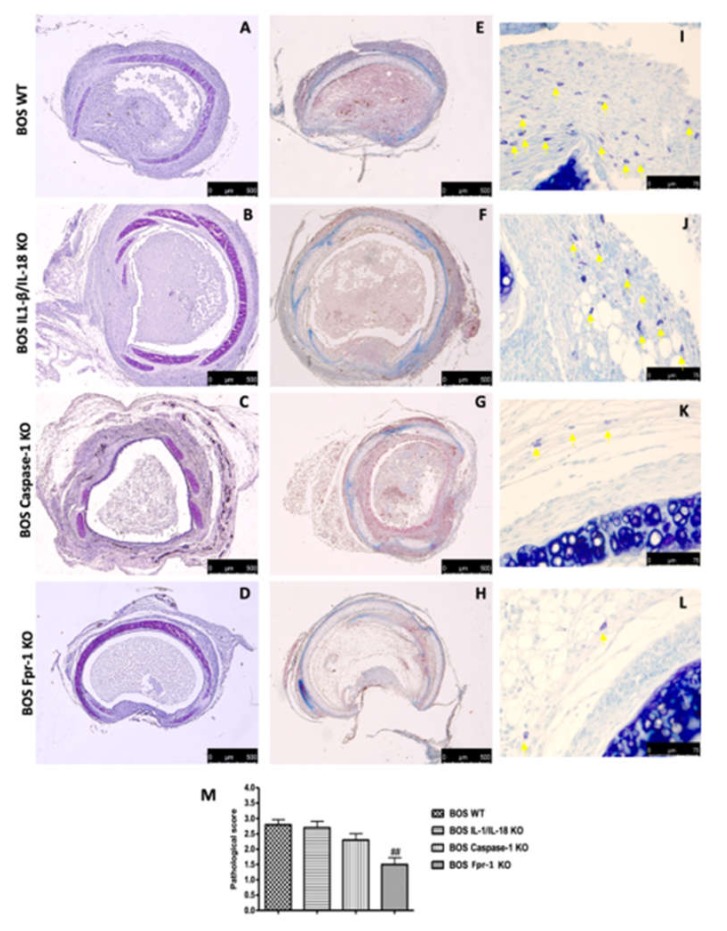
Figure 1.
Histopatology evaluation and mast cell density in IL-1β/IL-18 KO, Casp-1 KO, and Fpr-1 KO: Histological evaluation of tracheal transplantation: wild-type (WT) (A), IL-1β/IL-18 KO (B), Casp-1 KO (C), Fpr-1 KO (D). Masson trichrome staining of the graft: WT (E), IL-1β/IL-18 KO (F), Casp-1 KO (G), Fpr-1 KO (H). Evaluation of mast cell degranulation by toluidine blue: WT (I), IL-1β/IL-18 KO (J), Casp-1 KO (K), Fpr-1 KO (L). Histopathologic score (M). For histological analyses, n = 5 animals from each group were employed. A p-value less than 0.05 was considered significant. ## p < 0.01 versus the WT group.

