Figure 6.
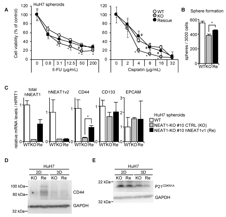
Phenotype rescue of NEAT1 deficiency in spheroids by hNEAT1v1 overexpression. (A) Viability of spheroids of HuH7 (white circles, WT), HuH7 NEAT1-KO #10 cells expressing mock (gray circles, KO) or hNEAT1v1 (black circles, Rescue) in the presence of 5-FU (left) or cisplatin (right). * p < 0.05 vs. WT; # p < 0.05 KO vs. Re, Turkey’s test (n = 4). (B) Spheroid formation ability of HuH7 cells (white columns, WT), and HuH7 NEAT1-KO #10 cells expressing mock (gray column, KO) or hNEAT1v1 (black column, Re). * p < 0.05, KO vs. Re; Tukey–Kramer’s test (n = 4). (C) mRNA expression levels relative to HPRT1 in spheroids of parental HuH7 cells (white columns, WT), and HuH7 NEAT1-KO #10 cells expressing mock (gray columns, KO) or hNEAT1v1 (black columns, Re). * p < 0.05, KO spheroids vs. Re spheroids; Tukey–Kramer’s test. (n = 3–4). (D,E) CD44 (D) and P21CDKN1A (E) protein expressions in monolayer cells (2D) and in spheroids (3D) of HuH7 NEAT1-KO #10 cells expressing mock (KO) or hNEAT1v1 (Re). GAPDH was used as an internal control.

