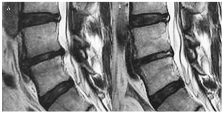
Figure 2.
A: Degenerative disc disease with Pfirrmann grade III disc showing an inhomogeneous structure, and an unclear distinction of nucleus and annulus and type 1 Modic changes with intermediate signal intensity in the T2 image with slightly decreased disc height and disc bulge. B: After radiofrequency ablation of the disc, sinuvertebral nerve, and basivertebral nerve, there is shrinkage of the degenerative disc and there is an increase in the signal of Modic changes in the adjacent vertebra body. However, with the current imaging technique, there are limitations in quantifying the effects of these end plate changes objectively. Further development in this area of assessment would be beneficial to assess treatment effects on the end plate and disc in early DDD (figure reproduced with permission courtesy of Kim et al. [12]).

