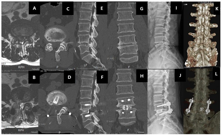
Figure 7.
Right-sided L4/5 recurrent disc herniation after previous open laminotomy and discectomy 2 years ago, the patient underwent uniportal endoscopic transforaminal lumbar interbody fusion of right L4/5(ETLIF). A shows the recurrence of disc herniation with facet arthritis in the MRI axial cut of L4/5. B shows the same axial cut with left transforaminal lumbar interbody fusion showing resection of the facet joint and cage in an optimal position. C and D show the pre and post-operative status of the right facet. Note the previous right L4 laminotomy in C, the facet was resected, and the cage introduced in an optimal position as shown in D. E and F show the sagittal view of the pre and pos-operative status. Note the increase in the foraminal height and intervertebral height as a result of the right L4/5 ETLIF. G and H show the increase in the coronal disc height pre and post-operatively in right L4/5 ETLIF. I and J show the pre and post-operative standing neutral XR with the L4/5 interbody cage and standard posterior inserted pedicle screws in L4 and L5. K and L show the 3D reconstruction of pre and post-operative right L4/5 ETLIF.

