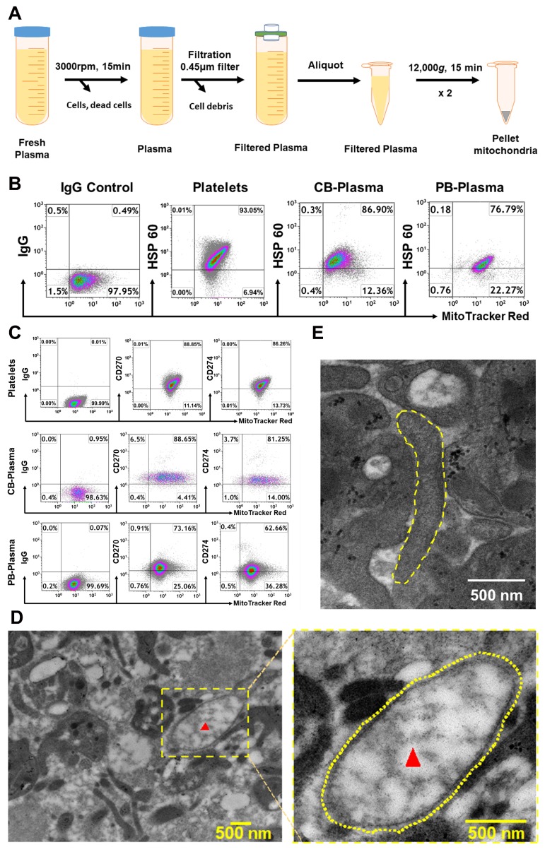
Figure 1.
Characterization of mitochondria in human blood. (A) Outline the protocol for an isolation of mitochondria from plasma. (B) Flow cytometry data show the plasma-derived mitochondria positive for specific mitochondrial markers (MitoTracker Deep Red) and heat shock protein (HSP) 60 from the plasma of human cord blood (CB, N = 4) and adult peripheral blood (PB, N = 4). Mitochondria isolated from the plasma of peripheral blood (N = 4) served as positive control. (C) Expression of immune tolerance-associated markers CD270 and CD274 on human CB (N = 8) and PB (N = 9) plasma-derived mitochondria respectively. Mitochondria isolated from peripheral blood-derived platelets served as positive control. Isotype-matched IgGs served as negative control. (D) Electron microscopy demonstrating the free mitochondria (red arrow, left) in the plasma of adult human blood, with a high magnification (yellow dashed circle, right). (E) Electron microscopy show a free elongated mitochondrion (yellow dashed circle) in adult human blood plasma.

