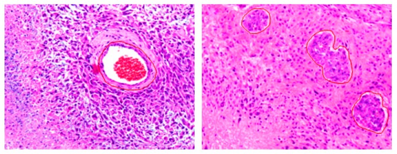
Figure 4.
Glioblastoma (grade-IV) tumors. Left: An island of viable tumor cells encircling the blood vessels in a large necrotic focus. Right: Tumor is experiencing endothelial proliferation. The image shows glomeruloid vessels and endothelial multilayering as a result of endothelial hyperplasia. These changes are driven by Vascular Endothelial Growth Factor.secreted by the tumor in response to hypoxia. The images were acquired using 20× magnification factor.

