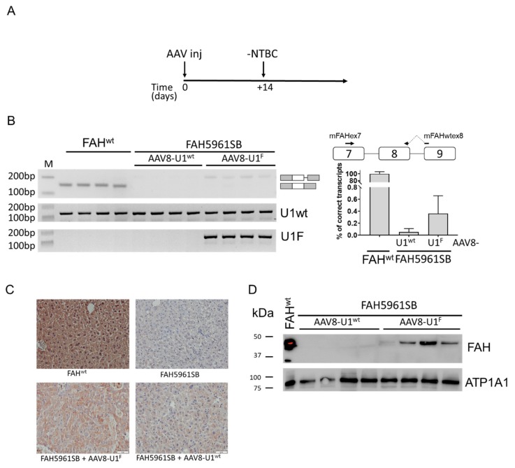
Figure 2.
The compensatory U1F partially rescues FAH expression in vivo. (A) Schematic representation of the protocol designed to perform the experiments in mice and exploiting the AAV8-mediated delivery of the U1snRNAs. Mice, kept on NTBC in their drinking water, were injected with 1*1013 vg/kg of body weight of AAV8-U1F or AAV8-U1wt and transferred to normal drinking water without NTBC (-NTBC) 14 days later (+14). (B) FAH splicing pattern profiles in mouse livers, together with the schematic representation of transcripts, reported on the right side. The quantification of correctly spliced FAH transcript by qPCR is indicated on the right, with the schematic organization of FAH gene region and the exploited primers (on top). Results are reported as percentage of correctly spliced transcripts (mean ± SD). (C) Immunohistochemical analysis of FAH expression in liver sections of wild type (FAHwt) and FAH5961SB mice, either untreated or treated with AAV8-U1wt or AAV8-U1F. Representative examples of liver sections stained with a specific anti-FAH antibody (brown). Images are taken at 20× magnification. Scale bar, 50 µm. (D) Western blotting analysis through a specific anti-FAH antibody in liver homogenates of wild type (FAHwt) and FAH5961SB mice treated with AAV8-U1wt or AAV8-U1F. The mouse ATPase Na+/K+ Transporting Subunit Alpha 1 (ATP1A1) was exploited as load control. The protein marker, reporting the molecular size of bands, is reported on the left.

