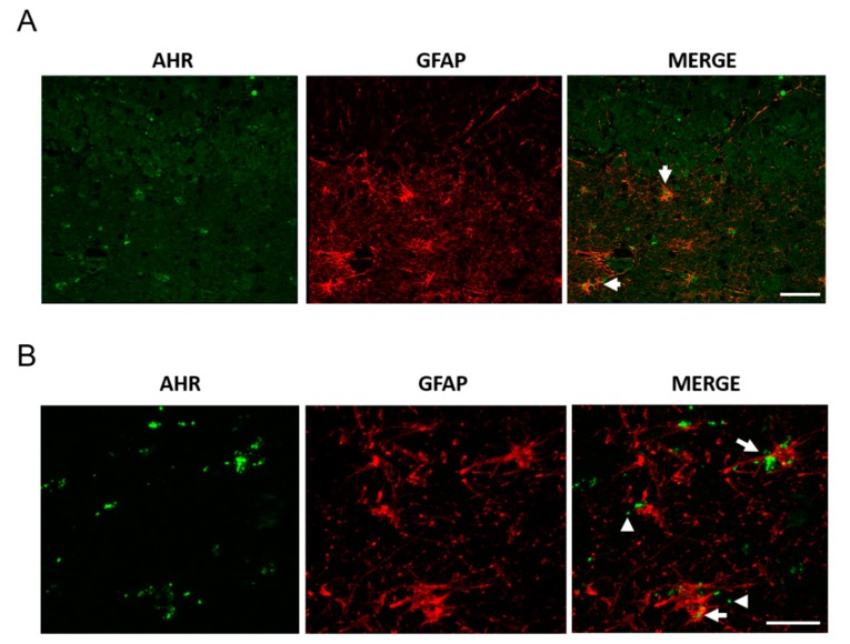
Figure 4.
AHR and GFAP colocalization in post-mortem tissue. We determined AHR and GFAP expression by immunostaining and found AHR in astrocytes, apparently as microvesicles. Representative images from a 94 year-old female donor (A, scale bar = 50 μm) and a 100 year-old Alzheimer’s disease (AD) female donor (B, scale bar = 20 μm). The arrows show the AHR staining in astrocytes. The arrowheads show AHR with the appearance of microvesicles. We analyzed samples from three donors.

