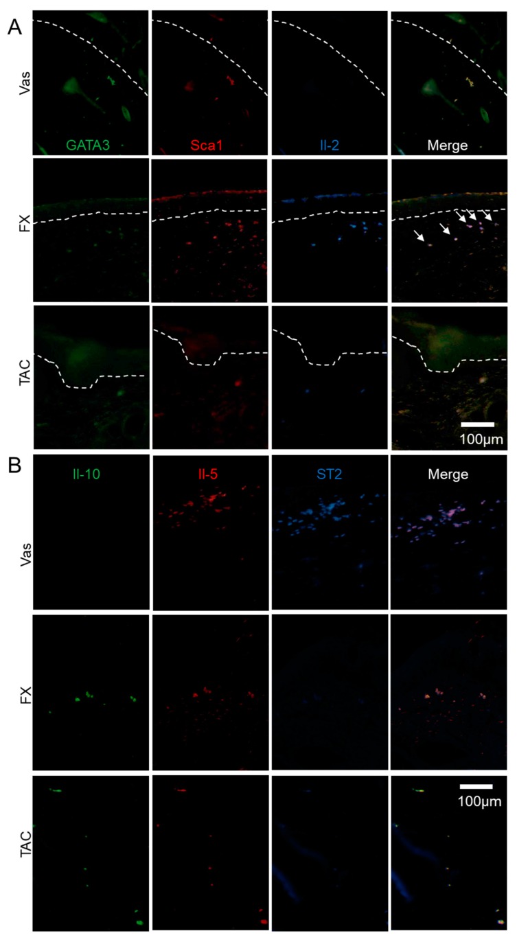
Figure 5.
The localization of ILC2 and ILCreg modulated by FX or TAC. FX stimulated Il-2+GATA3+Sca1+ ILCreg. Il-2+GATA3+Sca1+ ILCreg dominantly expressed in FX-treated Nc/Nga mice (A) and Il-5+ST2+ ILC2 in the Vas treated group (B). Sections were reacted with respective fluorescence-labelled antibodies. The area between the dotted lines indicates the epidermal layer. Arrows indicates typical GATA3lowIl-2high cells (A). Panel (B) is focused on the dermis.

