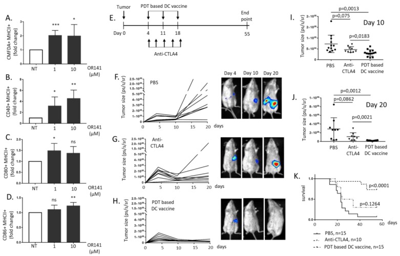
Figure 2.
Photodynamic therapy (PDT)-based dendritic cell (DC) vaccination leads to potent mesothelioma growth inhibitory effects. Naïve DCs were exposed to lysates from OR141-killed Ab1 mesothelioma cells for 18 h. (A) The phagocytotic activity was evaluated based on the increased incorporation of CMFDA-labeled Ab1 in CD11c+ DC (identified by flow cytometry) (n = 3, * p < 0.05, *** p < 0.001). (B–D) The extent of CD40+/MHCII+ (B), CD80+/MHCII+ (C) and CD86+/MHCII+. (D) The DC populations, as determined by flow cytometry (n = 3, * p < 0.05, ** p < 0.01; ns, non-significant). (E) A schematic representation of the drug regimens used, namely PDT-based DC vaccination and anti-CTLA4 immunotherapy. (F–K) The BALB/c mice were injected with 105 the luciferase-expressing Ab1 cell line (Ab1-luc) intraperitoneally (i.p.) and, at day four, were separated into three groups to evaluate the mesothelioma growth inhibitory effects of phosphate-buffered saline (PBS) (F), anti-CTLA4 antibodies (G) and the PDT-based DC vaccine (H); representative bioluminescence measurements are shown for each condition. The extent of the tumor size at day 10 (I) and day 20 (J) in the different conditions and corresponding Kaplan–Meier survival curves (K) (n = 10–15 mice).

