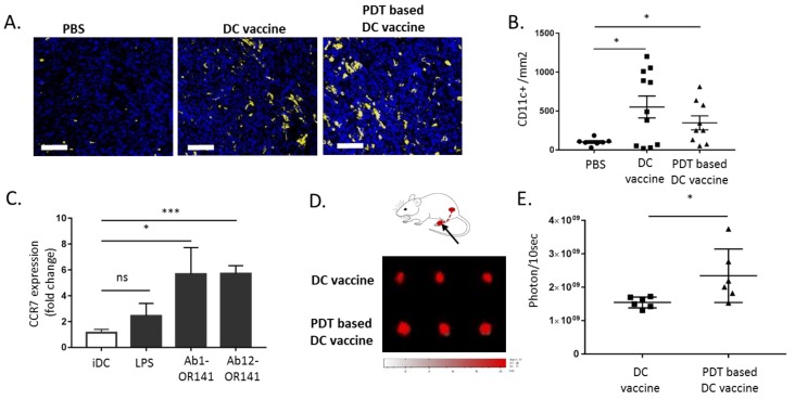
Figure 7.
DC motility towards lymph nodes is increased upon priming with PDT-killed mesothelioma cells. (A,B) The tumor-bearing BALB/c mice were injected with PBS, DC vaccine or PDT-based vaccine and sacrificed six days later for tumor immunostaining. (A) The representative CD11c+ staining images (scale bar = 50 µm) and (B) quantification (* p < 0.05, n = 8–10). (C) The DCs were exposed to lysates from OR141-killed Ab1 and Ab12 cells or to LPS and the extent of CCR7 surface expression was determined by flow cytometry (* p < 0.05, *** p < 0.001, n = 3). (D,E) The migration of DC, pre-labeled with VivoTrack 680 NIR, from the mouse footpad to the popliteal lymph nodes. (D) A schematic representation and representative fluorescence pictures of popliteal lymph nodes (PLN) collected three days post-injection from mice treated either with the DC vaccine or PDT-based DC vaccine. (E) The quantification of the extent of fluorescence detected in the PLN (* p < 0.05, n = 6).

