Figure 1.
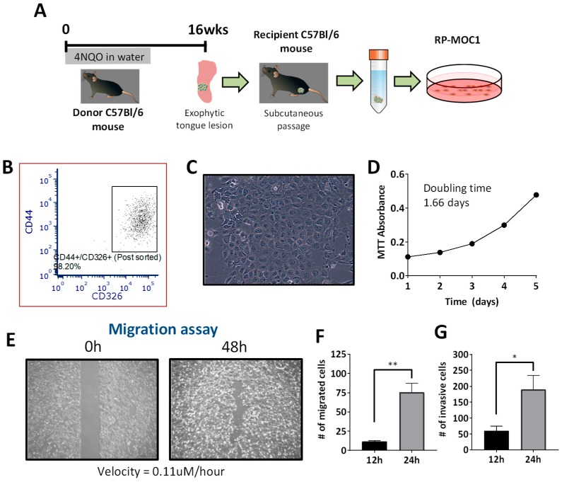
Establishment and in vitro behavior of the RP-MOC1 OSCC model. (A) Schematic shows workflow for generation of the RP-MOC1 cell line. Donor tongue tissue from a C57BL/6NCr mouse exposed to 4NQO in the drinking water for 16 weeks was initially transplanted subcutaneously to establish an allograft. The established tumor was subsequently excised, digested with collagenase and seeded in a petri dish. (B) Flow cytometry was used to sort cells co-expressing the stem cell marker (CD44) and epithelial cell adhesion molecule, EpCAM (CD326). (C) Microscopic image of RP-MOC1 showing characteristic cobblestone morphology with anisocytosis and cellular pleomorphism. Image was acquired at 20× magnification. (D) Growth curve of RP-MOC1 cells over a 5-day period. The doubling time for this cell line based on MTT was determined to be 37.6 ± 2.4 h. (E) Wound healing assay. Images of RP-MOC1 cells at 0 h and at 48 h are shown. The image at 48 h illustrates near complete wound closure due to migration of tumor cells. Images were acquired at 4× magnification. Scale bar: 500 μm. Bar graphs showing increase in the number of migrated cells (F) and invasive cells (G) between 12 h to 24 h. Data is reported as mean ± SEM and from two independent experiments. (ns, no significance; * p < 0.05; ** p < 0.01).

