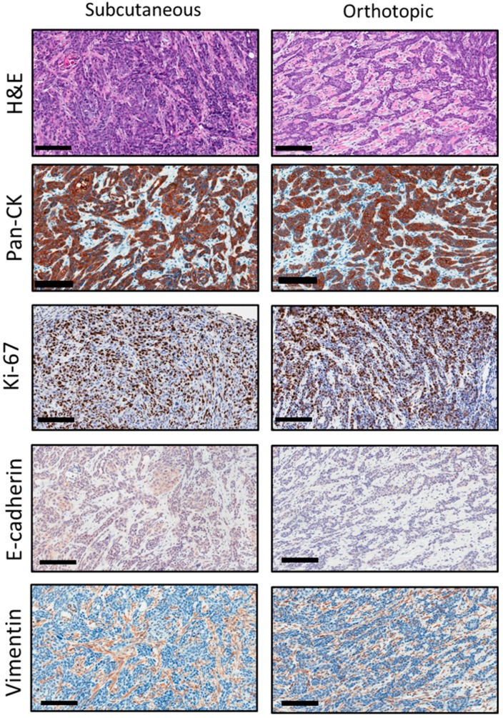
Figure 3.
Histopathologic credentialing of RP-MOC1 tumors. Photomicrographs of histology and immunohistochemical staining of tumor sections from the initial subcutaneous tumor and the subsequent orthotopic tumor are shown. Hematoxylin and eosin (H&E) stained sections showed invasive keratinizing moderately differentiated squamous cell carcinoma. Image was acquired at 20× magnification. Scale bar: 100 μm. Immunostaining for cytokeratin (Pan-CK) and Ki-67 showed positive staining on the membrane and nuclear staining of the epithelial cells. Immunostaining for E-cadherin and vimentin showed negative staining on the membrane and cytoplasm stain of the epithelial cells. Images were acquired at 20× magnification. Scale bar: 100 μm.

