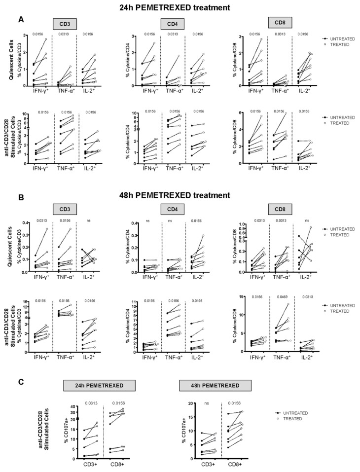
Figure 5.
In vitro pemetrexed treatment improved T-cell responses. (A) PBMCs from healthy donors (n = 7) were stimulated for 24 h with anti-CD3/anti-CD28 antibodies in the presence or absence of 100 nM pemetrexed. This was followed by co-staining for CD3, CD4, or CD8 plus IFN-γ, TNF-α, or IL-2 to determine T-cell function by flow cytometry. Upper and lower plots show results from unstimulated and stimulated PBMCs, respectively, with and without 24 h pemetrexed treatment. Data are presented as cytokine-positive, CD3+/CD4+/CD8+ T-cells detected in treated and untreated cultures, white and black dots, respectively. (B) PBMCs from healthy donors (n = 7) were stimulated for 48 h as in (A). (C) PBMCs from healthy donors (n = 6) were stimulated for 24 and 48 h as in (A) together with CD107a antibody to evaluate the cytotoxic potential by flow cytometry. Data are presented as CD107a-positive, CD3+, and CD8+ T-cells detected in treated and untreated cultures, white and black dots, respectively. (A–C) All data were analyzed with the Kolmogorov–Smirnov test to confirm or exclude a normal distribution. A statistical analysis was done by two-tailed Wilcoxon matched-pairs test and p-values are indicated.

