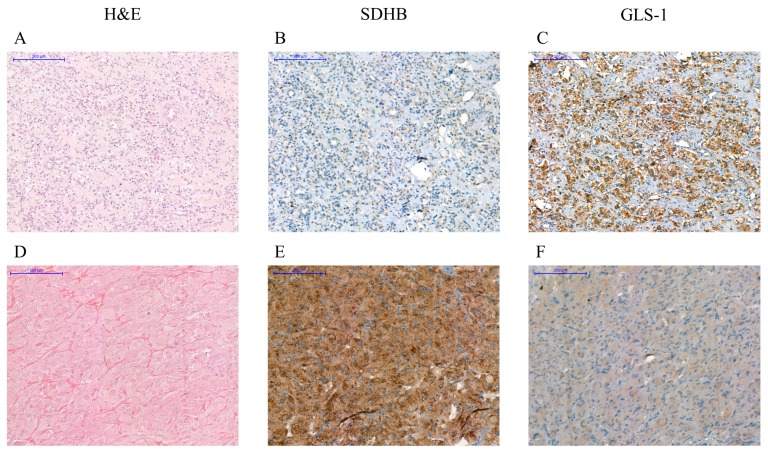
Figure 6.
Immunohistochemistry: Immunostaining with antibodies against SDHB and GLS-1 of a paraganglioma associated with SDHB p. mutation (A–C) and a RET p.C634W-associated pheochromocytoma (D–F). Lack of SDHB staining in SDHB mutated tumors (B) and strong GLS-1 signal was detected in malignant SDHB-associated tumor (C). Lack of GLS-1 positive cells can be observed in RET-associated benign pheochromocytoma (F). Scale bar = 200 µm. H&E: hematoxylin and eosin staining. SDHB: succinate dehydrogenase subunit B staining. GLS-1: glutaminase-1 staining.

