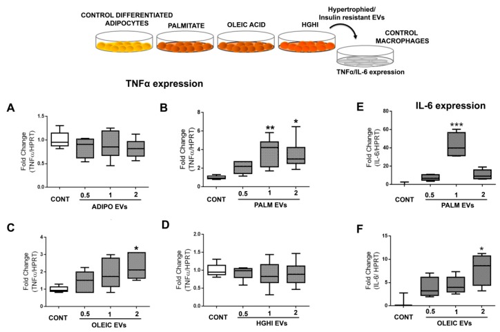
Figure 6.
EVs shed by hypertrophied adipocytes exert inflammation in macrophages. The effect of vesicles from lipid hypertrophied and HGHI adipocytes at different concentrations (physiological (1), half (0.5) and double doses (2)) over control non-inflamed macrophages was assayed by real time PCR expression of TNFα (A–D) and IL-6 (E–F). Box graphs are shown as fold change towards control adipocytes without any stimulus corrected by HPRT (at least 4 independent experiments; n = 3 replicates/experiment). Differences were assayed by one-way Anova-Kruskall Wallis test followed by Dunn´s multiple comparison test (p ≤ 0.05 was considered statistically significant: * p < 0.05, ** p < 0.01, and *** p < 0.001).

