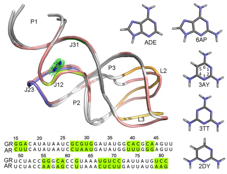
Figure 1.
The superimposition of structures and comparison of sequences for the guanine (GR) and adenine riboswitches (AR). The structures of riboswitches are shown in cartoon representation. The structures with different sequences of two purine riboswitches are colored in salmon color for GR, AR is colored with each secondary structure and labeled. The ligand in the binding pocket is shown using a stick-and-dot representation. Five ligands are also shown in stick representation.

