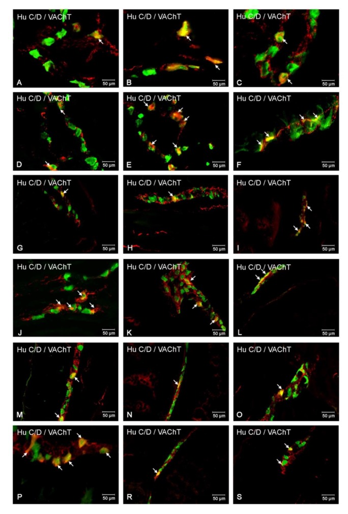
Figure 10.
Immunofluorescent staining presenting VAChT immunoreactivity in cell bodies in the particular intramural plexuses in the small intestine in both control and experimental groups. All photographs have been created by the digital superimposition of two color channels; Hu C/D-positive—used here as a pan-neuronal marker (green), and VAChT-positive (red). The arrow shows perikaryon containing both examined substances. Myenteric plexus of the duodenum (A,D), jejunum (B,E), and ileum (C,F) under physiological condition (A–C) and after streptozotocin administration (D–F). Outer submucosal plexus of the duodenum (G,J), jejunum (H,K), and ileum (I,L) under physiological condition (G-I) and after streptozotocin administration (J–L). Inner submucosal plexus of the duodenum (M,P), jejunum (N,R), and ileum (O,S) under physiological condition (M–O) and after streptozotocin administration (P–S).

