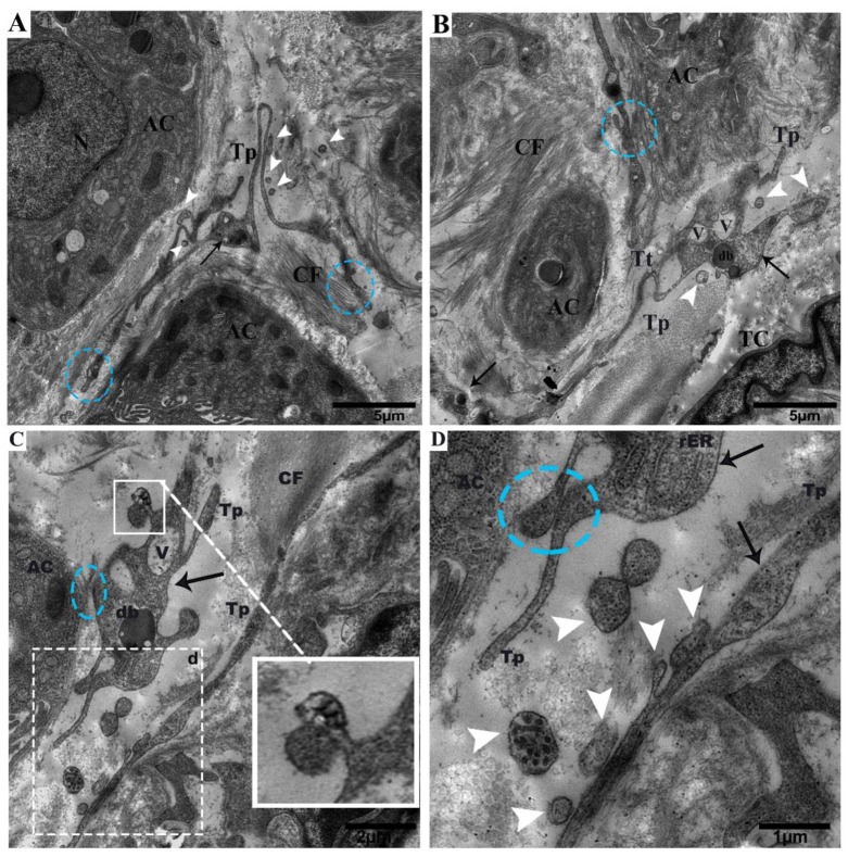
Figure 2.
Tps within the connective tissue budding and shedding microvesicles and rER in podoms, in between gland cells, making close connections with the centroacinus and neighboring cells. (A) Long rope-like Tps with a convoluted process forming junctions and close connections with collagen fibers (blue circle). The podoms have mitochondria and vesicles (black arrow). Shed vesicles within connective tissue (white arrowheads). (B) TCs have heterochromatin peripheral nuclei. Tps form a tortuous process in close proximity to the centroacinus, as well as close connections with gland cells and collagen fibers (blue circle). The podom contains vesicles and dense bodies (black arrows). Shed microvesicles from the podomic area within the connective tissue. Tps form a long process. (C) Vesicles and dense bodies (black arrow), and a close connection with the acinus (blue circle). A magnified view of budding extra cellular vesicles from the podom (white square). White circled area d in (C) inset into (D), respectively; in the podom rough endoplasmic reticulum (black arrow), close connections with podoms to podoms and the acinus can be seen (blue circle). Numerous shedding microvesicles within the connective tissue (white arrowheads) can be seen in (D). TC, telocyte; CF, collagen fibers; Tp, telopodes; Tt, tortuous process; db, dense body; V, vesicles; AC, acinus. Scale bar = 5 µm in (A,B); scale bar 2 µm in (C); scale bar = 1 µm in (D).

