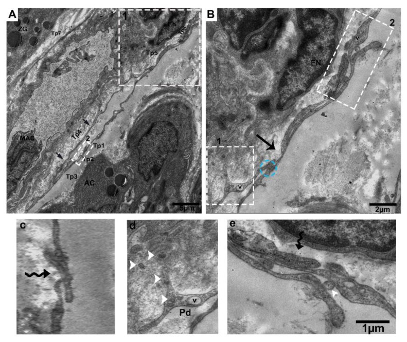
Figure 4.
Tps inside and surrounding the interlobular duct with different patterns of long and short overlapping podoms, and podomers within the connective tissue between the acinus. (A) Tps forming a long, convoluted process with many overlapping twists (black arrowheads), and the inside of the interlobular duct has podoms with vesicles. Square-marked areas in (A) inset into (B,C), respectively; (B) short overlap and punctate contact (blue circle) and gap junctions, and short overlap (black arrow). (c) Hook joint and short overlap with tight junctions between the three Tps. Square-marked areas in (B) inset into (d,e), respectively; (d) shed/extracellular vesicles within the space of the interlobular duct from the podom (white arrowheads). (e) A short overlap between podoms to podoms and podomers and shed vesicles (white arrowheads), in closeness to the endothelial cells. Tp, telopodes; MAC, macrophage; AC, acinus; E, endothelial; ZG, zymogen granules; V, vesicles; Pd, podoms. Scale bar = 5 µm in (A); scale bar = 2 µm in (B); scale bar = 1 µm in (e).

