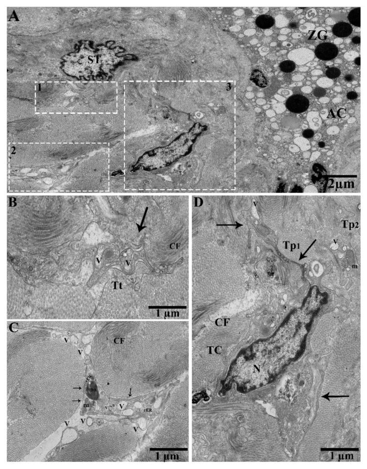
Figure 5.
In between gland cells, small blood capillaries, and numerous shed/extracellular vesicles from the podoms in the connective tissue in the vicinity of collagen fibers. (A) TEM photograph in the pancreas showing the acinar cells, zymogen granules, and stellate cells. Square-marked areas 1, 2, and 3 in (A) are enlarged in (B,C,D), respectively. (B) Tortuous process and vesicles (black arrow). (C) Podoms containing numerous extracellular/shed vesicles, scattered in the vicinity of collagen fibers, and rough endoplasmic reticulum, mitochondria, and dense bodies are present. (D) A typical TC, which contains heterochromatin in their periphery, forming Tp1 vesicles (arrows). Tp2 contains mitochondria and vesicles in the podoms. N, nucleus; ZG, zymogen granules; AC, acinus; ST, stellate cell; Tt, tortuous process; V, vesicles; m, mitochondria; rER, rough endoplasmic reticulum; DB, dense body; CF, collagen fibers. Scale bar = 2 µm in (A); scale bar =1 µm in (B–D).

