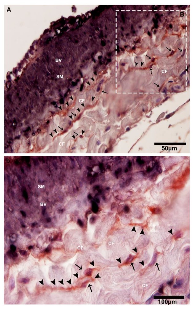
Figure 9.

CD34-positive and α-SMA-positive telocytes around the large blood vessels in the connective tissue. (A) The square-marked area B in (A) is enlarged in (b). Immunohistochemistry (IHC) analysis shows at lower magnifications the CD34-positive and α-SMA-positive cells adjacent to large blood vessels. At high magnifications, CD34 staining has strong positivity with dilated portions, whereas α-SMA stains positive for smooth muscle layers and blood vessels. Higher and lower magnifications of IHC (two anti-bodies—CD34 and α-SMA) were used. The square-marked area B in (A) is enlarged in (b). The telocytes and their nuclei can be seen (black arrows), showing long cellular projection telopodes (black arrows head) and displaying thin and thick (podoms and podomers) segments in the vicinity of collagen fibers and adjacent to the blood vessels. BV, blood vessels; CF, collagen fibers; SM, smooth muscle. Scale bar = 50 µm in (A); scale bar = 100 µm in (B).
