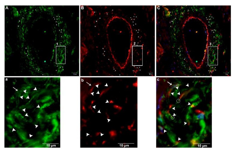
Figure 13.
Immunofluorescence-positive telocytes in the connective tissue surrounding large blood vessels. Immunofluorescence CD34+ is shown in green in (A), Vimentin+ in red in (B), and merged in (C), and labeled. Tps were abundantly distributed around the large blood vessels (arrowheads). The square-marked areas 1, 2, and 3 in (A,B,C) are enlarged in (a,b,c), respectively. TCs with Tps and long processes close to the other telopodes can be seen. (c) Podom, marked as a bright spot (circle), the nucleus (arrow), and the processes (arrowheads) are all visible. Scale bar = 20 µm in (A–C); scale bar = 20 µm in (a–c).

