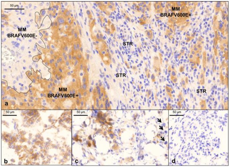
Figure 3.
IHC examples obtained from melanoma patients. (a) Note the intratumoral heterogeneity of the FFPE tissue sample. The BRAF V600E-mutated melanoma cells display no immunoexpression of the mutated protein (left of the dashed line); whereas strong immunoexpression of the mutated BRAF protein is evident to the right of the dashed line. Stromal inflammatory cells (STR) are the negative, internal controls for staining. (b) Homogenous patterns of expression are shown in cryosections of fresh frozen BRAF V600E-mutated melanoma samples. (c) Heterogeneous patterns of expression are shown in cryosections of fresh frozen BRAF V600E-mutated melanoma samples (arrows indicate melanoma cells that are negative for the mutated protein). (d) A fresh frozen wild-type melanoma sample is the negative control for the IHC method.

