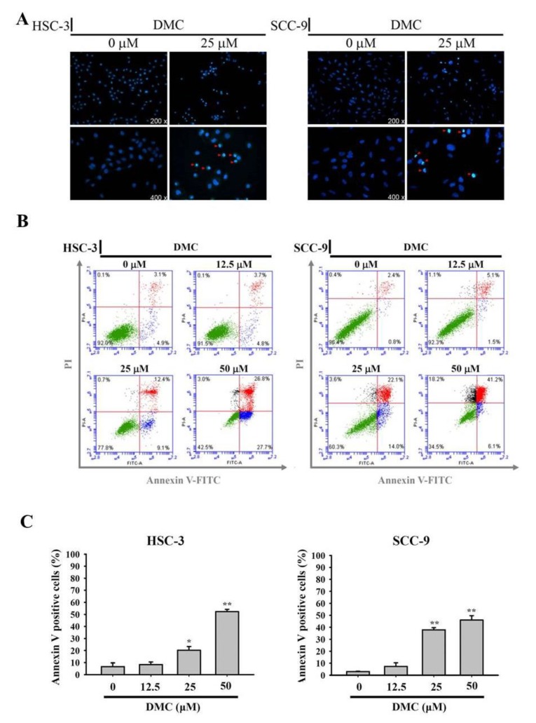
Figure 2.
Demethoxycurcumin (DMC) induces apoptotic cell death in oral squamous cell carcinoma (OSCC) cells. (A) After 24 h DMC (25 μM) treatment of SCC-9 and HSC-3 cells, the morphological characteristics of apoptosis were analyzed by fluorescence microscopy after Hoechst 33342 staining. The red arrows indicated the nuclear fragmentation and condensation which served as apoptosis indicators. (B,C) Quantitative analysis of cell apoptosis by Annexin-V and propidium iodide (PI) double-staining flow cytometry in SCC-9 and HSC-3 cells treated with DMC (12.5–50 μM) or the vehicle for 24 h. One representative example of both cells is displayed in B. Values represent the mean ± SD of three independent experiments (C). * p < 0.05, ** p < 0.01, compared to the vehicle group.

