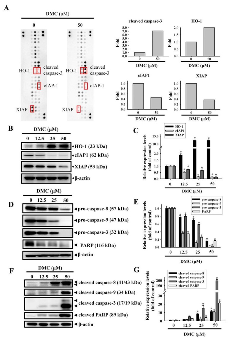
Figure 3.
High-throughput screening of apoptosis-related proteins modulated by demethoxycurcumin (DMC) in oral squamous cell carcinoma (OSCC) cells. (A, left panel) Representative images of the apoptotic protein array (R&D System) are shown for vehicle- and DMC-treated HSC-3 cells. (A, right panel) Proteins involved in apoptosis and regulatory pathways were quantitated using a densitometer and are represented as multiples of change compared to the controls. (B–G) HSC-3 cells were treated with indicated concentrations of DMC for 24 h, and a Western blot analysis was used to detect the expression levels of heme oxygenase (HO)-1, cellular inhibitor of apoptosis 1 (cIAP1), X-chromosome-linked IAP (XIAP), pro- and cleaved caspases-3, -8, and -9, and poly(ADP-ribose) polymerase (PARP) (B,D,F). The β-actin protein levels were used to adjust the quantitative results of these protein levels and expressed as multiples of induction beyond each respective control. Values are presented as mean ± SD from three independent experiments. * p < 0.05, compared to the vehicle group (C,E,G).

