Figure 2.
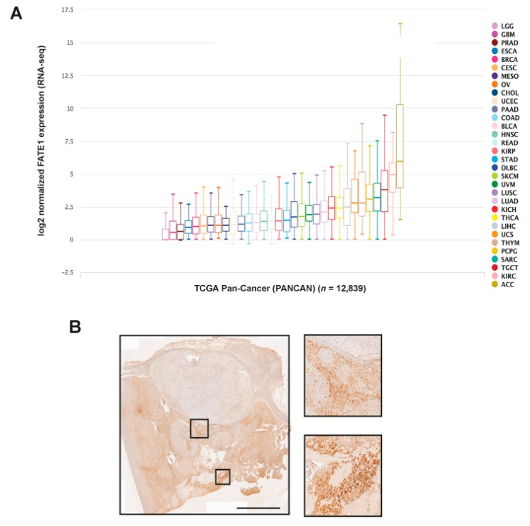
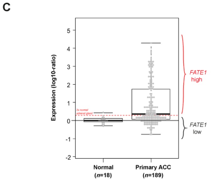
FATE1 expression in adult ACC. (A) FATE1 mRNA expression in cancers of the TCGA PANCAN dataset. 12,839 cases in total were analyzed using the Xena browser (https://xenabrowser.net). Tumor types are color-coded. ACC, adrenocortical carcinoma; KIRC, kidney clear cell carcinoma; TGCT, testicular germ cell cancer; SARC, sarcoma; PCPG, pheocromocytoma-paraganglioma; THYM, thymoma; UCS, uterine carcinosarcoma; LIHC, liver hepatocellular carcinoma; THCA, thyroid carcinoma; KICH, kidney chromophobe cell carcinoma; LUAD, lung adenocarcinoma; LUSC, lung squamous cell carcinoma; UVM, uveal melanoma; SKCM, skin cutaneous melanoma; DLBC, diffuse large B-cell lymphoma; STAD, stomach adenocarcinoma; KIRP, kidney papillary cell carcinoma; READ, rectum adenocarcinoma; HNSC, head and neck squamous cell carcinoma; BLCA, bladder urothelial carcinoma; COAD, colon adenocarcinoma; PAAD, pancreatic adenocarcinoma; UCEC, uterine corpus endometrial carcinoma; CHOL, cholangiocarcinoma; OV, ovarian serous cystadenocarcinoma; MESO, mesothelioma; CESC, cervical squamous cell carcinoma and endocervical adenocarcinoma; BRCA, breast invasive carcinoma; ESCA, esophageal carcinoma; PRAD, prostate adenocarcinoma; GBM, glioblastoma multiforme; LCG, brain lower grade glioma. (B) FATE1 IHC staining in an ACC from an adult patient. Some tumor areas display intense FATE1 staining, other nodules appear to express very little FATE1. Two groups of FATE1 positive cells are shown at higher magnification. Scale bar, 5 mm. (C) FATE1 mRNA expression across 189 adult ACC and 18 normal adrenal samples. Histogram of distribution of FATE1 mRNA expression levels. The red horizontal line defines the “FATE1-low” and “FATE1-high” classes.


