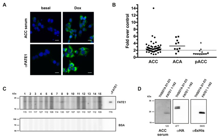
Figure 5.
IF, ELISA and Western blots for circulating anti-FATE1 antibodies. (A) IF: mitochondrial staining by an ACC serum selectively in Dox-treated, but not untreated, H295R/TR N-Flag FATE1 cells. Anti-FATE1 monoclonal antibody staining is shown as a control. Scale bar, 20 μm. (B) ELISA: graph showing results for ACC, ACA and pediatric ACC (pACC). Samples generating OD450 signals higher than two-fold negative controls (dotted line) were considered as positive. (C) Western blot: sera reactivities were tested against recombinant FATE1 (1–162) and BSA as negative control. A representative blot is shown using samples from patients 1 to 15 in Table 4. Band signals (values under each lane in the FATE1 immunoblot) are expressed as a percentage over the local background. (D) Specificity control for Western blot: a serum from a patient with ACC recognizes recombinant FATE1 (aa. 1–162) but not another recombinant human autoantigen of similar molecular weight (thrombospondin type-1 domains 1 to 3 of THSD7A [31]). Recombinant proteins were detected with anti-hemagglutinin (HA; THSD7A) and anti-6xHis-Tag (FATE1) antibodies, respectively. Band signals (values under each lane) are expressed as a percentage over the local background. Original uncropped blots are shown in Figure S1A,B.

