Figure 3.
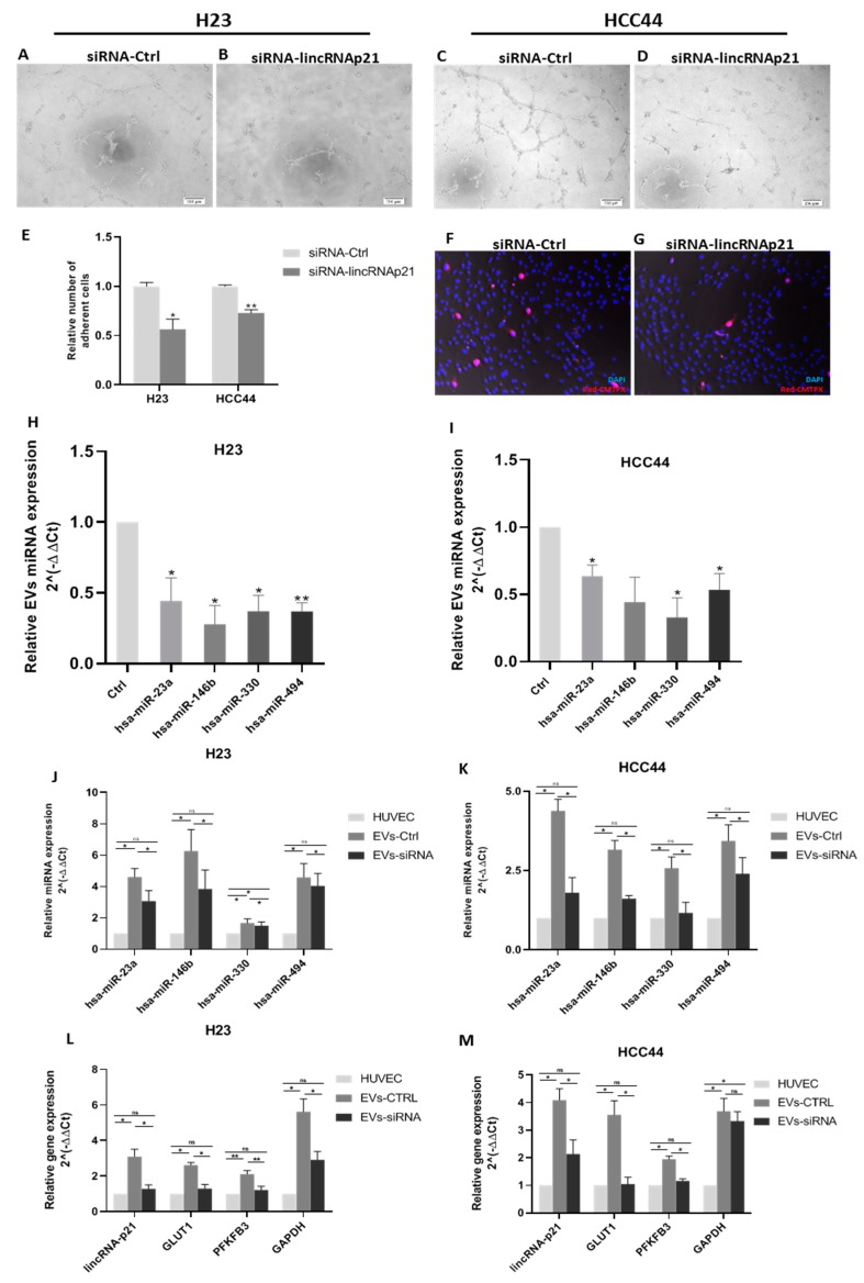
Functional in vitro assays to evaluate the modulation of angiogenesis and endothelial cell permeability by EV treatment. (A–D) Tube formation assay using human umbilical vein endothelial cells (HUVECs) treated with control EVs in H23 (A) and HCC44 (C) and lincRNA-p21-silenced EVs in H23 (B) and HCC44 (D). (E) Quantification of relative number of adherent cells in a cell adhesion assay using a monolayer of HUVECs treated for 12 h with control EVs or lincRNA-p21-silenced EVs. (F,G) A representative image of HCC44 cell adhesion assay. Blue staining using DAPI indicates nucleus and HCC44 cells were stained with cell tracker in red (20× magnification). (H,I) lincRNA-p21 silencing reduced the EV levels of miR-23a, miR-146b, miR-330, and miR-494 in both cell lines. (J,K) EV conditioning increases the expression of miR-23a, miR-146b, miR-330, and miR-494 in HUVECs. (L,M) EV conditioning increases the expression of lincRNA-p21, GLUT1, PFKFB3, and GAPDH in HUVECs. * p < 0.05; ** p < 0.01.

