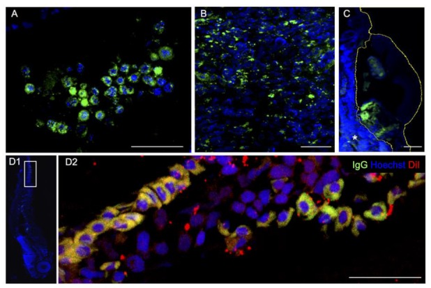
Figure 6.

Anti-Human IgG immunohistochemistry (green). (A) HCT 116 cancer cell lines two days post injection into the zebrafish yolk. Patient’s pancreatic tumor before (B) and after xenotransplantation, 2 dpi (C). (D1,D2) Patient’s colon cancer cells spread throughout the vasculature reaching the zebrafish tail. Human cells are labeled with DiI (red) and anti-IgG (green). D1 is a magnification (90° rotation) of D2. Scale bar 50 μm.
