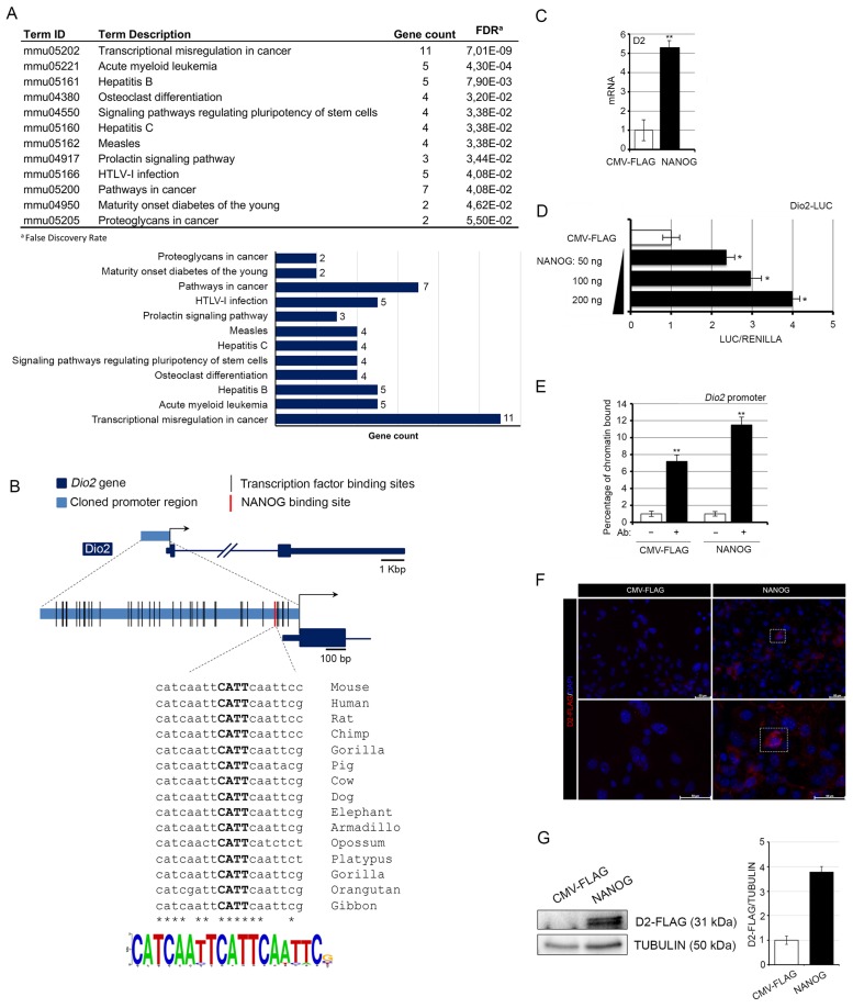
Figure 1.
NANOG positively regulates type 2 deiodinase (D2) transcription. (A) KEGG pathway clusters generated by the in silico analysis of the Dio2 promoter region. (B) Schematic localization of transcription factors and of the NANOG binding site within the Dio2 promoter region; conservation and Logo representation of the NANOG binding motif. (C) D2 mRNA expression was measured by real-time PCR in basal cell carcinoma (BCC) cells transiently transfected with a NANOG-expressing vector or the CMV-FLAG plasmid (control). (D) BCC cells were transiently transfected with the Dio2–LUC promoter and with increasing amounts of the NANOG plasmid. Cells were harvested 48 h after transfection and analyzed for luciferase activity. CMV-Renilla was co-transfected as an internal control. The results are shown as means ± SD of the LUC/Renilla ratio from at least three separate experiments, performed in triplicate; * p < 0.05, ** p < 0.01. (E) Chromatin immunoprecipitation assay was performed in BCC cells. Immunoprecipitation of chromatin using the anti-NANOG antibody revealed that the Dio2 gene is a direct target of NANOG. (F) D2-FLAG expression levels were measured by immunofluorescence analysis of mouse primary keratinocytes from D2-Flag mice transfected with the NANOG plasmid or the CMV-FLAG plasmid. Magnification 10× and 20×; scale bars represents 50 μm. (G) Western Blot analysis of D2 expression in the same cells as in F.

