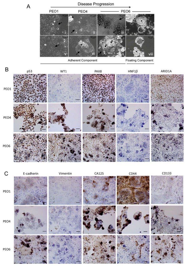
Figure 1.
(A) PEO1, PEO4, and PEO6 cells were cultured for 10 days in media with 10% serum, including insulin and antibiotics. Arrows: adherent cells. Arrowheads: 3D foci. Squares: irregular multicellular structures. Asterisks: spheroidal multicellular structures. (B) Expression of biomarkers of HGSOC in cultured PEO1/4/6 cells. (C) Expression of markers of epithelial-mesenchymal transition and of stem cell plasticity in PEO1/4/6 cells. Scale bars = 50 μm.

