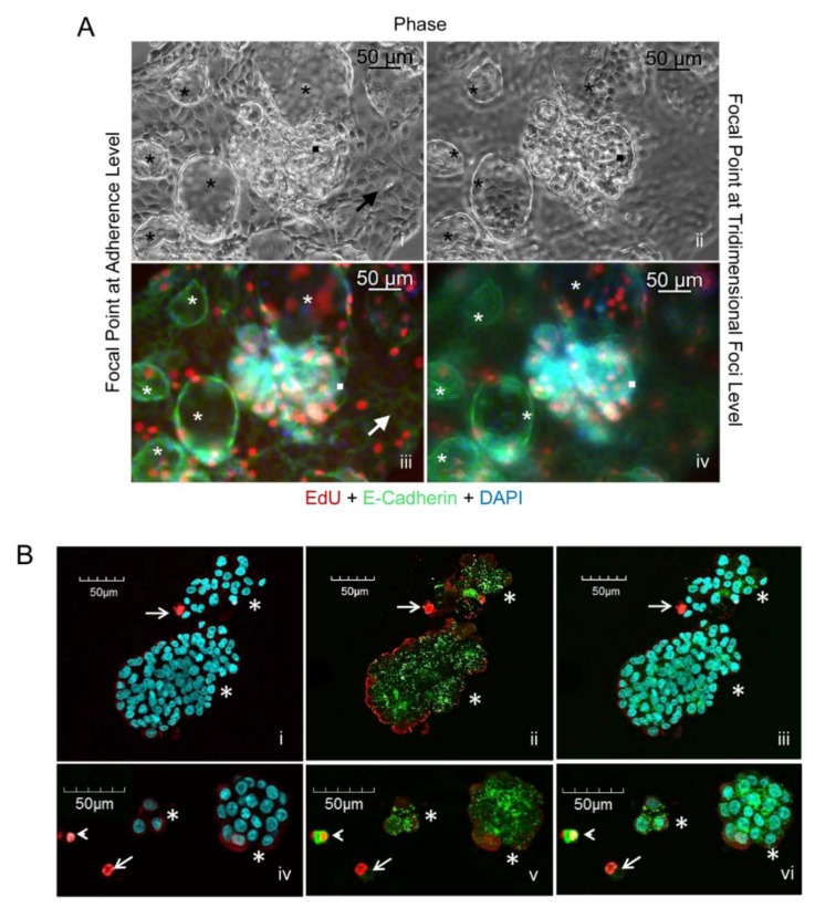
Figure 2.
(A) Phase contrast imaging at focal [i] and 3D planes [ii] of PEO6 cells displaying a cellular monolayer (arrow), 3D irregular multicellular structures (squares), and spheroidal multicellular structures (asterisks). EdU incorporation (red fluorescence) and E-cadherin expression (green fluorescence) are shown at the focal plane [iii] and at the 3D plane [iv]. Blue, DAPI, depicting nuclei. (B) Live/dead® viability/cytotoxicity assay of PEO6 cells arranged as multicellular structures as stained with DAPI [i, iv], calcein AM and EthD-1 [ii, v], and their overlay [iii, vi]. Asterisks denote multicellular structures mostly alive as assessed by their green calcein AM staining. Arrowheads denote dying cells (yellow), whereas arrows identify dead cells (red).

