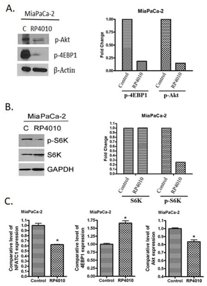
Figure 2.
RP4010 inhibited calcium-regulated Akt/mTOR and NFAT signaling. (A and B) MiaPaCa-2 cells were grown overnight in 100-mm petri dishes to nearly 50% confluence. The cells were then treated on the following day with RP4010 (10 µM) for 72 h. Protein extraction, determination of protein concentration, SDS-PAGE, and Western blot were performed as described in the Methods (n = 1). β-actin and GAPDH were used as loading controls. The expression of marker proteins was indicated as fold change relative to the control, and the quantitative analysis of mean pixel density of the blots was performed using ImageJ software. (C) MiaPaCa-2 cells were exposed to RP4010 (10 µM) for 48 h. At the end of the treatment period, RNA was isolated and RT-qPCR was performed as described in Methods (* p < 0.05).

