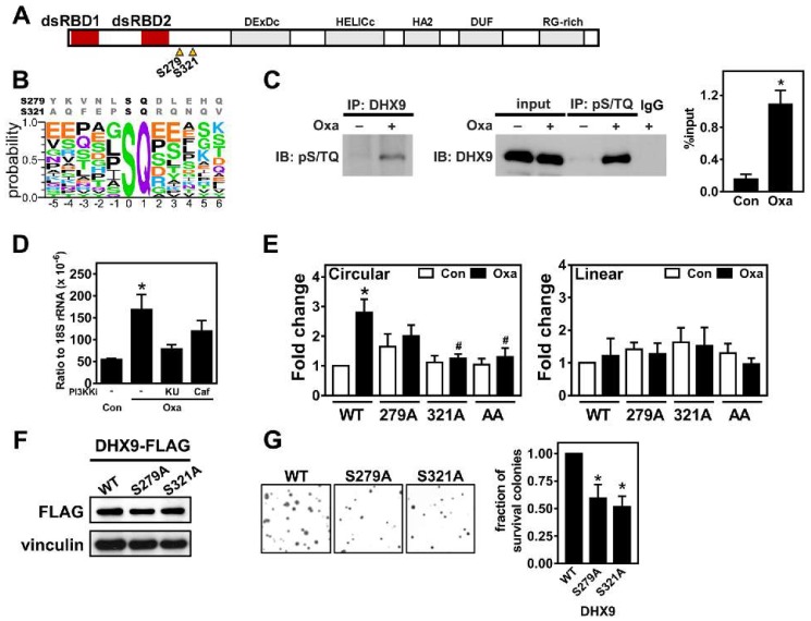
Figure 4.
Oxaliplatin-induced DHX9 phosphorylation promotes circRNA expression. (A) Illustration of DHX9 domains. Triangles: Potential phosphorylation sites at serine 279 and 321. (B) Sequences around predicted PI3KK phosphorylation sites in DHX9 (top) compared to known sequences (Bottom image was generated by using WebLogo [31]. Y-axis: probability for each amino acid at the corresponding position and X-axis: position relative to phosphorylated serine). (C) Representative images of an immunoblot (IB) using an antibody recognizing the pS/TQ motif from immunoprecipitated (IP)-DHX9 (left). In a reciprocal fashion, representative images of a DHX9 immunoblot from immunoprecipitated proteins containing the pS/TQ motif (middle). Levels of phospho-DHX9 are shown as % of input (right). Input: lysates without IP. Oxa: cells treated without (–) or with (+) oxaliplatin (1 μg/mL) for 8 h. IgG: IP using rabbit nonimmune immunoglobulin. (D) Levels of circCCRC66 in the cells treated with indicated compounds for 24 h. Oxa: oxaliplatin (1 μg/mL), KU: KU-55933 (10 μM) and Caf: caffeine (1 mM). * p < 0.05, significantly different to control group. (E) The levels of the circular transcript and mRNA (linear transcript) of CCDC66 determined by quantitative PCR in oxaliplatin-treated HCT116 cells transfected with denoted DHX9 constructs (WT: wildtype; nonphosphorable versions: serine residue at 279 or 321 mutated to alanine or combination of both (AA)). * p < 0.05, significantly different to WT/con. # p < 0.05, significantly different to WT/oxa. (F) The representative images of immunoblots for HCT116 transfected with the DHX9 expressing vector. Vinculin serves as the loading control. (G) Similar to (E), representative images for a clonogenic assay performed in cells transfected with indicated DHX9 constructs and treated with 1-µg/mL oxaliplatin for more than 7 days. * p < 0.05, significantly different to WT.

