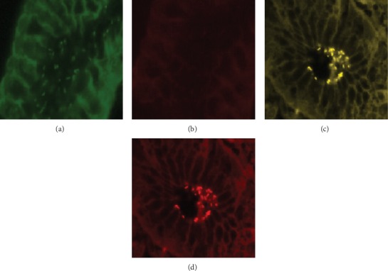
Figure 2.

Specific detection of clarithromycin-sensitive and clarithromycin-resistant H. pylori isolates in gastric biopsy specimens by FISH. (a) Identification of bacteria in green, due to hybridization with probe Hpy-1–FITC. (b) Visualization of the same microscopic field in red cannel, indicating strain is clarithromycin-sensitive H. pylori. (c) Demonstration of clarithromycin-resistant H. pylori in the biopsy specimen of a patient by simultaneous application of probe Hpy-1–FITC and the mixture of probes ClaR1-Cy3, ClaR2-Cy3, and ClaR3-Cy3; bacteria are visible in yellow (mixed color of green and red). (d) Visualization of the same microscopic field in red cannel, due to hybridization with probe ClaR1-Cy3, ClaR2-Cy3, or ClaR3-Cy3.
